|
Reference Information
• Do Nuclear Power Plants Cause Cancer?
Some Applications of Nuclear Technologies
• Food and Agriculture Applications
• Nuclear Medicine Applications
• Non-Destructive Testing Applications
Food and Agriculture Applications
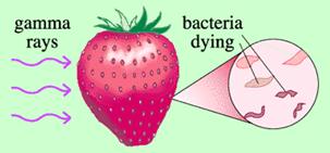
One of the popular applications of nuclear technology is food irradiation. Food irradiation can eliminate bacteria, insects and parasites that can cause food-borne diseases, such as salmonella, trichinosis and cholera. In addition, irradiation can slow down spoilage and increase the shelf life of food.
The irradiation process exposes food to gamma rays from cobalt-60 which is a radioisotope of cobalt. Gamma rays are a form of electromagnetic energy, just like radio waves, microwaves, X-rays and even light. Gamma rays have the ability to penetrate well into a food. Machine-generated X-rays have similar properties. More recently, electron beams (e-beams) have become available as a source of ionizing energy in the USA and other countries. Like X-rays, e-beams are machine-generated using ordinary electricity and can be powered on and off at the touch of a switch. E-beams offer extremely rapid and cost-effective processing, but in some cases sacrifice penetration depth depending on product density. Treatment of food using either X-rays or electron beams are occasionally referred to as “electronic pasteurization” or “electronic irradiation” methods because they are derived from electricity.
Regardless of the source of ionizing energy, the food is treated by exposing it to the energy source for a precise time period. In the case of e-beam, food is irradiated in just a few seconds, while it takes gamma and X-rays considerably longer. The food is never in contact with the energy source; the ionizing energy merely penetrates into the food but does not stay in the food. It takes very little energy to destroy harmful bacteria. At these levels there is no significant increase in temperature or change in composition. Irradiation does not make food radioactive nor does it leave any residues.
Irradiation does not change the food any more than canning or freezing. All known methods of food processing and even storing food at room temperature for a few hours after harvesting can lower the content of some nutrients, such as vitamins. At low doses of radiation, nutrient losses are either not measurable or, if they can be measured, are not significant. At the higher doses used to extend shelf-life or control harmful bacteria, nutritional losses are less than or about the same as cooking and freezing. Independent scientific committees in USA, Denmark, Sweden, United Kingdom and Canada also have reaffirmed the safety of food irradiation. In addition, food irradiation has received official international endorsement from the World Health Organizations and the International Atomic Energy Agency. In the United States, the U.S. Food and Drug Administration has approved the use of irradiation for fruits, vegetables, pork, poultry, red meat and spices. In addition, more than 40 countries have also approved the use of radiation to help preserve nearly 40 different varieties of food.
In agriculture, radiation had eradicated approximately 10 species of pest insects in wide areas, preventing agricultural catastrophes. These pests included the Mediterranean fruit fly and the screwworm fly. The technique used for eradicating the aforementioned insects is the sterile insect technique which is a method of biological control, whereby millions of insects sterilized with radiation, are released. The released insects are normally male as it is the female that causes the damage, usually by laying eggs in the crop, or, in the case of mosquitoes, taking a bloodmeal from humans. The sterile males compete with the wild males for female insects. If a female mates with a sterile male then it will have no offspring, thus reducing the next generation's population. Repeated release of insects can eventually wipe out a population, though it is often more useful to consider controlling the population rather than eradicating it.
Agricultural researchers also use radiation to:
- develop hundreds of varieties of hardier, more disease-resistant crops—including peanuts, tomatoes, onions, rice, soybeans and barley;
- improve the nutritional value of some crops, as well as improve their baking or melting qualities or reduce their cooking time;
- pinpoint where illnesses strike animals, allowing the breeding of disease-resistant livestock;
- show how plants absorb fertilizer, helping researchers to learn when to apply fertilizer, and how much to use; this prevents overuse, thus reducing a major source of soil and water pollution.
References:
- Robert J. Woods, “Food irradiation”, Endeavour, Volume 18, Issue 3, 1994, Pages 104-108
- Johannes Friedrich Die, “Will irradiation enhance or reduce food safety?”, Food Policy, Volume 18, Issue 2, April 1993, Pages 143-151
- L. H. Wedekind, “Food irradiation in Asia and the Pacific”, Food Policy, Volume 11, Issue 4, November 1986, Pages 285-288
- S. V. Nerpin, V. M. Prokhorov and V. N. Savin, “Use of isotopes and nuclear radiation in agricultural research “, Atomic Energy, Publisher Springer New York, ISSN 1063-4258 (Print) 1573-8205 (Online), Issue Volume 26, Number 2, February, 1969, pp. 161–165
- http://www.osti.gov/bridge/servlets/purl/840065-Jhd7iT/native/840065.pdf
- http://www.iaea.org/nafa/d5/index.html
- http://physics.isu.edu/radinf/food.htm
- http://www.nei.org/howitworks/foodandagriculture/
Go to Top
Nuclear Medicine Applications
In nuclear medicine, medical professionals inject a tiny amount of a radioisotope—a chemical element that produces radiation—into a patient’s body. A specific organ picks up the radioisotope, enabling a special camera to take a detailed picture of how that organ is functioning. In modern nuclear medicine, PET and SPECT are two widely used imaging techniques.
PET stands for Positron Emission Tomography. It is a nuclear medicine tomographic imaging technique which produces a three-dimensional image or map of functional processes in the body. The system detects pairs of gamma rays emitted indirectly by a positron-emitting radionuclide (tracer), which is injected into a patient’s body. Images of tracer concentration in 3-dimensional space within the body are then reconstructed by computer analysis. The patient’s body will never come in contact with scanner itself. PET scans can be used to measure metabolic activity and molecular function by using a radioactive glucose injection such as F-18 FDG (Fluorine-18 Fluorodeoxyglucose). Although all cells use glucose as an energy source, cancer cells grow faster than normal healthy cells and they use glucose at much higher rate than normal cells. This is the basis of imaging with F-18 FDG for cancer detection in PET scan.
SPECT stands for Single Photon Emission Computed Tomography. SPECT imaging is performed by using a gamma camera to acquire multiple 2-D images (also called projections), from multiple angles. A computer is then used to apply a tomographic reconstruction algorithm to the multiple projections, yielding a 3-D dataset. This dataset may then be manipulated to show thin slices along any chosen axis of the body. SPECT is similar to PET in its use of radioactive tracer material and detection of gamma rays. In contrast with PET, however, the tracer used in SPECT emits gamma radiation that is measured directly, whereas PET tracer emits positrons which annihilate with electrons up to a few millimeters away, causing two gamma photons to be emitted in opposite directions. A PET scanner detects these emissions "coincident" in time, which provides more radiation event localization information and thus higher resolution images than SPECT (which has about 1 cm). SPECT scans, however, are significantly less expensive.
Radionuclides used in nuclear medicine are mostly artificial ones. They are primarily produced in a reactor or cyclotron and supplied by commercial companies to individual nuclear medicine departments and institutions. On the other hand, some radionuclides, in particular short-lived ones, are available at any time due to the availability of appropriate radionuclide generators. By far the most important generator in nuclear medicine is the 99Mo/99mTc generator, which has led to the almost unlimited availability of 99mTc.
Technetium-99m is a metastable nuclear isomer of technetium-99, symbolized as 99mTc. The "m" indicates that this is a metastable nuclear isomer, i.e. it does not change into another element (transmutate) upon its "decay". It is a gamma ray emitting isotope used in radioactive isotope medical tests, for example as a radioactive tracer that medical equipment can detect in the body. It is well suited to the role because it emits readily detectable 140 keV gamma rays (these are about the same wavelength emitted by conventional X-ray diagnostic equipment), and its half-life for gamma emission is 6.01 hours (meaning that about 93.7% of it decays to 99Tc in 24 hours). The short half life of the isotope allows for scanning procedures which collect data rapidly, but keep total patient radiation exposure low.
These kinds of diagnostic procedures involve very small amounts of radioisotopes. In higher doses, radioisotopes also help treat disease. For example, radioactive iodine’s widespread use in therapy for thyroid cancer results in a lower recurrence rate than drug therapy. It also avoids potentially fatal side effects, such as the destruction of bone marrow.
Sealed sources of radiation placed inside the body, or radiation directed from external sources, are effective in treating various cancers. Nearly half of all cancer patients in the United States receive radiation treatment at some point in their therapy.
Hospitals also use radiation to sterilize materials, thus helping to prevent the spread of diseases. Exposing these materials to radiation does not make them radioactive.
References:
Go to Top
Non-Destructive Testing Applications
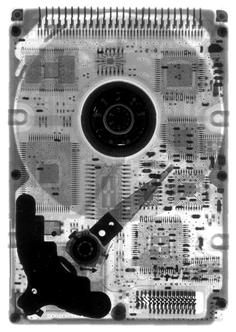
Non-Destructive Testing (NDT) is a term for describing a class of inspection methods for detecting defects in objects or structures without destroying the original specimens. Among the various NDT methods, radiography is one of the popular methods. Radiography uses X-rays and gamma-rays to produce an image of a specimen on film, showing any changes in thickness, defects (internal and external), and assembly details to ensure optimum quality in the operation.
X-rays and Gamma rays are electromagnetic radiation of exactly the same nature as light, but of much shorter wavelength. Wavelength of visible light is on the order of 6000 angstroms while the wavelength of x-rays is in the range of one angstrom and that of gamma rays is 0.0001 angstrom. This very short wavelength is what gives x-rays and gamma rays their power to penetrate materials that light cannot. This high penetration capability of the rays allows us to see the internal conditions of the object which cannot be seen from outside by naked eyes. If an x-ray or gamma ray source is placed on one side of a specimen and a photographic film on the other side, an image is obtained on the film of the thickness variations in the specimen. This is a well-established NDT technique and is widely used to detect internal flaws in weldments and castings and to check for mis-constructions in assemblies
X-rays are produced by an x-ray generator and is usually described by the electrical voltage across the x-ray tube. The higher the voltage, the greater is the penetrating power of the radiation; industrial x-ray equipment ranges from about 20KV to 20 MV and the most powerful equipments can be used to radiograph up to 500mm steel. Nearly all gamma-radiography is done with either cobalt-60 or iridium-192 sources. To obtain good definition images, it is desirable to have small-diameter radiation sources and the effective source size is in the range of 1 to 4mm diameter. After the radiographic film has been exposed, it has to be photographically processed (develop, wash, fix and dry) and is then placed on an illuminated screen for visual interpretation of the image. X-rays and Gamma-rays are dangerous and must be used either inside a protective enclosure or appropriate barriers and warning signals.
|
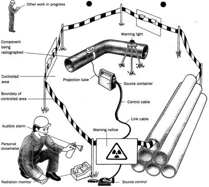
Site arrangement for carrying out radiography
|
References:
Go to Top
|